|
This is a young
female, age 34, who was operated for bilateral hydrosalpinx.
During the gross examination of the specimen, the left
fallopian tube was found completely obstructed by the presence
of a tumor like lesion, of relatively compact consistency and
grayish appearance.
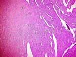
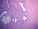
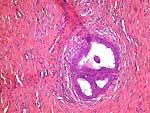
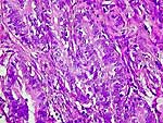
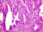
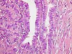
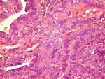
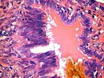
Microscopic examination of
several slides reveals the presence of a hyperplastic
papillary lesion, which in some places exhibits high grade
cytological atypia. These papillary formations are
lined by one or more rows of atypical epithelial cells
suggesting malignant infiltration.
The pictures shown here were
taken with magnifications of 4x, 10x, 20x and 40x, with a Nikon 400 Microscope,
using a Nikon Coolpix 900 Digital Camera.
We present eight images from
sections stained with hematoxylin & eosin.
Please reply to:
Konstadinos
Syllas, MD
|
Case
presented by:
Konstadinos
Syllas, MD
Pathologist
Toxicological
Laboratory
Aristotle's University of Thessaloniki
Ermou 36 str.,
546 23 Thessaloniki - GREECE Tel - fax : 031 - 262132
|